Day 1 :
Keynote Forum
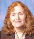
Heliah S. Oz
University of Kentucky Medical Center, KY USA
Keynote: Inflammatory diseases, postsurgical tissue adhesion complications, current practices and novel emerging targeted therapies
Time : 10:00-10:45

Biography:
Dr Helieh Oz has a DVM, MS (U. IL), PhD (U. MN) and clinical translational research certificate (U. KY Med Center). Dr Oz is an active member of American Association of Gastroenterology (AGA) and AGA Fellow (AGAF). She is an immuno-microbiologist/pathologist with expertise in inflammatory, infectious diseases, signal transduction, pathogenesis, innate/mucosal Immunity, micronutrient and drug discovery. Dr Oz has over 90 publications in the areas of chronic inflammatory disorders (pancreatitis, hepatitis, colitis, periodontitis), microbial and infectious diseases, signalling pathways and tissue repair. Dr Oz has served as Lead editor for special issues such as Gut Inflammatory, Infectious diseases and Nutrition (Mediators of Inflammation 2017); Nutrients, Infectious and Inflammatory Diseases (Nutrients 2017); Gastrointestinal Inflammation and Repair: Role of Microbiome, Infection, Nutrition (Gastroenterology Research Practice 2016), and co-editor for Parasitic infections in pediatric clinical practice (J Pediatric Infectious Disease 2016) and Chagas Disease Book, Intech Open Science 2017. Dr Oz serves as member of different editorial advisory board committees including Center of Excellence for Medical Research and Innovative Products, Walailak University and is an avid reviewer for several peer-reviewed journals
Abstract:
Tissue adhesion occurs following mechanical trauma or extensive inflammatory tissue injuries. Tissue adhesions remain as significant clinical challenges affecting millions of patients each year. Subsequent abdominal surgeries and wound healing, patients may be challenged with formation of postsurgical tissue adhesion (PSTA) complications as consequences of tissue repair. Approximately 65-97% of patients suffer from some types of PSTAs, when an organ surface is damaged due to inadvertent desiccation or trauma. During healing, tissues may firmly attach to the adjacent surfaces by the formation of fibrous scars. The mechanism of PSTA formation is similar to wound healing and response to implants. PSTAs can be asymptomatic, or followed by complications comprise of abdominal or pelvic pain, intestinal obstructions and infertility. Further, these adhesions form complex tissue barriers making subsequent surgical interventions costly and increasingly difficult if not life threatening. In addition to severe negative impact on quality of life, the annual financial expenditure related to PSTA exceeds $1.3 billion. Therefore, effective strategies for preventing PSTA formation remain significant clinical challenges.
In this keynote presentation, the PSTA formations and consequences will be discussed in patients, following in in vitro and in vivo models and the current therapeutic practices and their short comes. In addition, new and emerging strategies to speed wound healing such as utilization of fibrin-targeted PSTA prevention material (e.g. fibrin gel matrix) and nanocomposite-based, biodegradable tissue adhesives will be scrutinized in protecting against adhesion formations without interfering in tissue wound healing process or causing further complications.
Keynote Forum
Paul Lucas
New York Medical College, USA
Keynote: Multipotent Adult Stem Cells (MASCs): Differentiation and regeneration

Biography:
Paul Lucas earned his Ph.D. in Biochemistry from the University of Minnesota. He was a Postdoctoral Fellow with Dr. Arnold I. Caplan from 1986 to 1987. He was an Associate Professor of Surgery at Mercer University School of Medicine. From 1997, he has been Director of Orthopaedic Research and Associate Professor of Orthopaedic Surgery and Associate Professor of Pathology at New York Medical College, Valhalla NY. He is an inventor on 6 issued patents and 2 patents pending. His research has focused on adult stem cells and tissue regeneration.
Abstract:
Tissue regeneration in adult humans requires large numbers of cells. We are working with a unique population of adult stem cells: multipotent adult stem cells (MASCs). MASCs have properties which make them uniquely useful for tissue regeneration: 1) an apparently unlimited proliferation potential in vitro, 2) the ability to differentiate into phenotypes of all 3 dermal lineages, 3) ability to respond to local signals to differentiate to the tissues at the site and 4) do not elicit an immune rejection response.
MASCs have been isolated from embryonic chicks, adult rats, mice, rabbits, and humans. They have been isolated from skeletal muscle, bone marrow, fat, and skin. MASCs from all species and all tissues are isolated and cultured by the same protocol, and they all exhibit the same proliferative and differentiation behavior in vitro and response to local factors in vivo. Differentiated phenotypes observed in vitro include skeletal myotubes, chondrocytes, osteoblasts, adipocytes, smooth muscle cells, endothelial cells, cardiomyocytes, fibroblasts, astrocytes, neurons, oligodendrocytes, keratinocytes, hepatocytes, pancreatic islet cells, and epithelial cells. Rat MASCs have been taken to 300 cell doublings, mice to 600 cell doublings, and human to 100 cell doublings.
In vivo regeneration models where MASCs have been tested include meniscal defect in rabbits, cartilage defects in rabbits, femoral and calvarial bone defects in rats, dermal defects in rats, an open tibial defect in rats, and injection sub-q of human MASCs into young adult rats. In the open tibia defect (Fig 1), MASCs were observed to differentiate into 7 phenotypes: keratinocytes, hair follicle cells, gland cells, endothelial cells, smooth muscle cells, fibroblasts, and periosteum. When injected into young adult rats, human MASCs were observed to differentiate into endothelial, smooth muscle, and hair follicle keratinocytes.
MASCs have the potential to regenerate tissues and become a useful tool in regenerative medicine.
Keynote Forum
Jean-Marc Lemaitre
Saint Eloi Hospital, France
Keynote: Exploring strategies for cell rejuvenation and tissue regeneration: iPSC reprogramming as a unique opportunity to understand and cure aging
Time : 11:50-12:35

Biography:
Ph.D. in Molecular and cellular biology of development, senior scientist since 2014 and awarded in 2006 for an AVENIR INSERM Team program on aging, he is currently Deputy Director at the Institute of Regenerative Medicine and Biotherapy (IRMB), the leader of a INSERM research team and Director of a stem cell facility CHU (SAFE-iPSC). He was invited speaker in 48 national and international conferences and seminars in France and abroad in the last 5 years.
Abstract:
Many of the pathologies that could benefit from regenerative stem cell-based therapies are associated to aging. Emerging evidences indicates that adult stem cells exhibit functional shortcomings, including pronounced shifts in the types of mature effector cells produced as well as alterations in self-renewal capacity. Many intrinsic or extrinsic stress are able to accelerate the exhaustion of the proliferative capacity of stem cells or differentiated progenitors towards an ultimate senescence-like cell cycle arrest. Important and specific epigenetic modifications have been observed during this process, likely driving a specific gene expression « signature of cellular aging », and little is known about changes in large-scale genome organization during this aging process and or in different during senescence induced situations and its relationship with genetic instability. To further analyse this process and the relationship between replicative stress and chromatin reorganization, we followed the reorganization of chromatin dynamics features associated with senescence induction as well as the associated changes of the DNA replication program (Riviera-Mulia et al., 2017, Ogrunc et al., 2016, Prieur et al., 2016). To further understand the interplay between genetics and epigenetics in tissue aging and to unravel molecular barriers, preventing cell rejuvenation of the age-related cellular physiology, we developped reprogramming strategies of somatic cells into induced pluripotent stem cells (iPSCs) to erase the hallmarks of cellular aging. Although this strategy provides a unique opportunity to derive patient-specific stem cells with potential application in autologous tissue replacement, limitation was revealed for elderly individuals, due to senescence described as a barrier to reprogramming that could drive genetic instability. To overcome this barrier and improve tissue regeneration, we developed an optimized reprogramming strategy that caused efficient reversing of cellular senescence and aging through reprogramming towards pluripotency. We demonstrated that iPSCs derived from senescent and centenarian fibroblasts have reset all the hallmarks of cellular aging, as telomere size, gene expression profiles, oxidative stress and mitochondrial metabolism, and are undistinguishable from hESC. Finally, we further demonstrate that re-differentiation, led to rejuvenated cells with a reset cellular physiology maintaining genetic stability, defining a new paradigm for human cell rejuvenation (Milhavet and Lemaitre 2014, Venables et al., 2013, Lapasset et al. 2011). Then we applied this knowledge to develop iPSC models for premature aging syndromes with high risk of genetic instability, to further explore the relationship between pathological and physiological aging. We will present and discuss data concerning opportunities and limits of using the iPSC technology for modelling pathologies involving replication stress, leading to senescence and ageing and genetic instability (Riviera-Mulia et al., 2017, Bouckenheimer et al., 2016, Lemey et al., 2016, Besnard et al., 2012, 2014).
Keynote Forum
Tojan Rahhal
University of Missouri, USA
Keynote: Pulmonary delivery of butyrylcholinesterase as a model protein to the lung

Biography:
Dr. Tojan Rahhal is an Adjunct Assistant Professor in Bioengineering and the Director of Diversity and Outreach Initiatives at the University of Missouri-Columbia in the College of Engineering. Rahhal graduated from North Carolina State University with a BS in Biomedical Engineering. She went on to pursue a PhD in Pharmaceutical Sciences at the University of North Carolina at Chapel Hill (UNC-Ch), working in the lab of Dr. Joseph M. DeSimone. Rahhal’s research focused on Engineering PRINT Particles for Pulmonary Delivery of Therapeutics and examined the effect of particle parameters (size, shape, composition, and surface chemistry) on residence time, cellular interactions, and immune responses in the lungs. Her work addresses the need for more efficient delivery of active therapeutics/biologics using dry powders that allow for monodisperse aerosolization and accurate deposition in the lungs for treatment of pulmonary diseases. Rahhal’s work has been published in Molecular Pharmaceutics and Nanomedicine: Nanotechnology, Biology, and Medicine.
Abstract:
Pulmonary delivery has great potential for delivering biologics to the lung if the challenges of maintaining activity, stability, and ideal aerosol characteristics can be overcome. To study the interactions of a biologic in the lung, we chose butyrylcholinesterase (BuChE) as our model enzyme, which has application for use as a bioscavenger protecting against organophosphate exposure or for use with pseudocholinesterase deficient patients. In mice, orotracheal administration of free BuChE resulted in 72 h detection in the lungs and 48 h in the broncheoalveolar lavage fluid (BALF). Free BuChE administered to the lung of all mouse backgrounds (Nude, C57BL/6, and BALB/c) showed evidence of an acute cytokine (IL-6, TNF-α, MIP2, and KC) and cellular immune response that subsided within 48 h, indicating relatively safe administration of this non-native biologic. We then developed a formulation of BuChE using Particle Replication in Non-Wetting Templates (PRINT). Aerosol characterization demonstrated biologically active BuChE 1 μm cylindrical particles with a mass median aerodynamic diameter of 2.77 μm, indicative of promising airway deposition via dry powder inhalers (DPI). Furthermore, particulate BuChE delivered via dry powder insufflation showed residence time of 48 h in the lungs and BALF. The in vivo residence time, immune response, and safety of particulate BuChE delivered via a pulmonary route, along with the cascade impaction distribution of dry powder PRINT BuChE, showed promise in the ability to deliver active enzymes with ideal deposition characteristics. These findings provide evidence for the feasibility of optimizing the use of BuChE in the clinic; PRINT BuChE particles can be readily formulated for use in DPIs, providing a convenient and effective treatment option.
Keynote Forum
Fumio Arai
Kyushu University, Japan
Keynote: The novel mesenchymal stromal niche cell in the bone marrow endosteum

Biography:
Fumio Arai is a Professor of Department of stem cell biology and Medicine, Graduate School of Medical Sciences, Kyushu University. He has completed his Ph.D. at the age of 28 years from Meikai University and postdoctoral studies from Keio University School of Medicine. His research interest is in studying the mechanisms of the cell fate regulation of HSCs at the single cell level for the establishment of the system that can expand HSCs.
Abstract:
Hematopoietic stem cells (HSCs) are responsible for blood cell production throughout the lifetime of individuals. Interaction of HSCs with their supportive microenvironmental niche, which is composed of cellular components located around stem cells, facilitate the signaling networks that control the balance between self-renewal and differentiation. HSCs maintain a quiescent state in the bone marrow, where they anchor to specialized niches along the endosteum (the border between the bone and the BM) and in perivascular sites adjacent to the endothelium. The cells in the endosteal niche are a heterogeneous population regarding their degree of differentiation and accompanying functions. In this study, we further characterized the endosteal niche cell populations by using the single-cell gene expression analysis and identified a small subpopulation in ALCAM+Sca-1– osteoblastic cell fraction that expressed pluripotent stem cell markers. Furthermore, this subpopulation of ALCAM+Sca-1– cells specifically expressed Cdh2. Also, this newly identified subpopulation could differentiate into osteoblast, adipocyte, and chondrocyte, and showed the gene expression pattern that closes to ES cells rather than other bone marrow MSC populations. We also evaluated the function of ALCAM+Sca-1–Cdh2+ cells and found that have the potential to maintain the self-renewal activity of HSCs. These data suggest that ALCAM+Sca-1–Cdh2+ cells are mesenchymal stromal cells with niche cell activity for HSCs.
